2026
 |
71.-
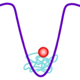
Single Nanoparticle Dynamics in Opto-Thermal Tweezers: Unveiling the Temporal Resolution of Depletion Force Trapping
J. Chen, R. Talla Kontchou, S. Rai, G. Baffou, S. Blaize, Q. Jiang, and J. Wenger*
ACS Nano , accepted (2026)
 Abstract:
Optothermal tweezers enable the manipulation of a wide range of nano-objects through optically induced depletion forces. Despite significant advances, the temporal dynamics of optothermal trapping remain elusive, as existing methodologies rely almost exclusively on time and ensemble averaging. Consequently, stable trapping cannot be distinguished from local transient accumulation, where the time-averaged concentration increases but particles exhibit rapid, dynamic motion in and out of the trap. Here we investigate optothermal trapping with single-nanoparticle-level analysis and sub-millisecond temporal resolution. Our data resolve the elusive dynamics of 40 nm polystyrene nanoparticles trapped within depletion force potentials in polyethylene glycol solutions, enabling to differentiate the conditions leading to extended trapping times from those leading to transient localization. Numerical simulations corroborate our experimental findings, elucidating how the interplay between thermophoresis and diffusiophoresis governs nanoparticle dynamics. These insights deepen our mechanistic understanding of optothermal trapping and unlock new opportunities for single-molecule studies, nanoscale assembly, and targeted drug delivery.
|
2025
 |
69.-
Surface modifications induced by the laser ablation of surface-bound microparticles at low to moderate fluence level in the ultraviolet
Alexandre Beaudier, Baptiste Marthy, Charles Bouyer, Romain Parreault, Guillaume Baffou, Jerome Neauport*
Optics Express 33, 6359 (2025)
 Abstract:
We investigate surface modifications induced by the ablation of fused glass and aluminum micro-particle contamination exposed to laser shots at a wavelength of 351 nm, with fluences ranging from 3.5 to 9.4 J/cm2. We establish a proportionality relationship between the size of the particle and the size of the crater created by the particle ablation on the substrate, which depends on the nature of the particle and the fluence level. Quadriwave lateral shearing interferometry (QLSI) microscopy is used to acquire high-resolution phase shift and amplitude maps of the surface modifications. We show that the combination of particle type and fluence level can lead to different amplitude and phase surface modifications. Diffraction modeling using these measurements evidences that aluminum particles can lead to up to 4.5x light intensification on the first centimeter after the surface, while glass particles exhibit a shallow intensification. We also show that for this type of particle there is a widely dispersed behavior at intermediate fluences in the 5 to 8 J/cm2 range. For a given fluence in this range, we experienced various phase/amplitude distributions in the damage sites, sometimes leading to strong intensifications.
|
2024
 |
68.-
Quantitative phase microscopies: accuracy comparison
P. C. Chaumet, P. Bon, G. Maire, A. Sentenac, G. Baffou*
Light: Science and Applications 13, 288 (2024)
Abstract:This article presents a thorough comparison of themain QPM techniques, focusing on their accuracy in terms of measurement precision and trueness. We focus on 8 techniques, namely digital holographic microscopy (DHM), cross-grating wavefront microscopy (CGM), which is based on QLSI (quadriwave lateral shearing interferometry), diffraction phase microscopy (DPM), differential phase-contrast (DPC) microscopy, phase-shifting interferometry (PSI) imaging, Fourier phase microscopy (FPM), spatial light interference microscopy (SLIM), and transport-of-intensity equation (TIE) imaging. For this purpose, we used a home-made numerical toolbox based on discrete dipole approximation (IF-DDA). This toolbox is designed to compute the electromagnetic field at the sample plane of a microscope, irrespective of the object's complexity or the illumination conditions. We upgraded this toolbox to enable it to model any type of QPM, and to take into account shot noise. In a nutshell, the results show that DHM and PSI are inherently free from artefacts and rather suffer from coherent noise; In CGM, DPC, DPM and TIE, there is a trade off between precision and trueness, which can be balanced by varying one experimental parameter; FPM and SLIM suffer from inherent artefacts that cannot be discarded experimentally in most cases, making the techniques not quantitative especially for large objects covering a large part of the field of view, such as eukaryotic cells.
|
2023
 |
66.-
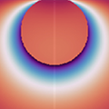
Quantitative Microscale Thermometry in Droplets Loaded with Gold Nanoparticles
L. Sixdenier,* G. Baffou, C. Tribet, E. Marie*
Journal of Physical Chemistry Letters 14, 11200-11207 (2023)
Abstract:Gold nanoparticles (AuNPs) are increasingly used for their thermoplasmonic properties, i.e. their ability to convert light energy into heat through plasmon resonance. However, measuring temperature gradients generated at the microscale by assemblies of AuNPs remains challenging, especially for random 3D distributions of AuNPs. Here, we introduce a label-free thermometry approach, combining quantitative wavefront microscopy and numerical simulations, to infer the heating power dissipated by a 3D model system consisting of emulsion microdroplets loaded with AuNPs. This approach gives access to the temperature reached in the droplets upon laser irradiation without the need for extrinsic calibration. These quantitative results are validated via the observation of the phase transition of a thermoresponsive polymer added in the droplets as an in situ thermal probe. This versatile thermometry approach is promising for non-invasive temperature measurements in various 3D microsystems involving AuNPs as colloidal heat sources, including photothermal drug delivery systems.
|
 |
65.-
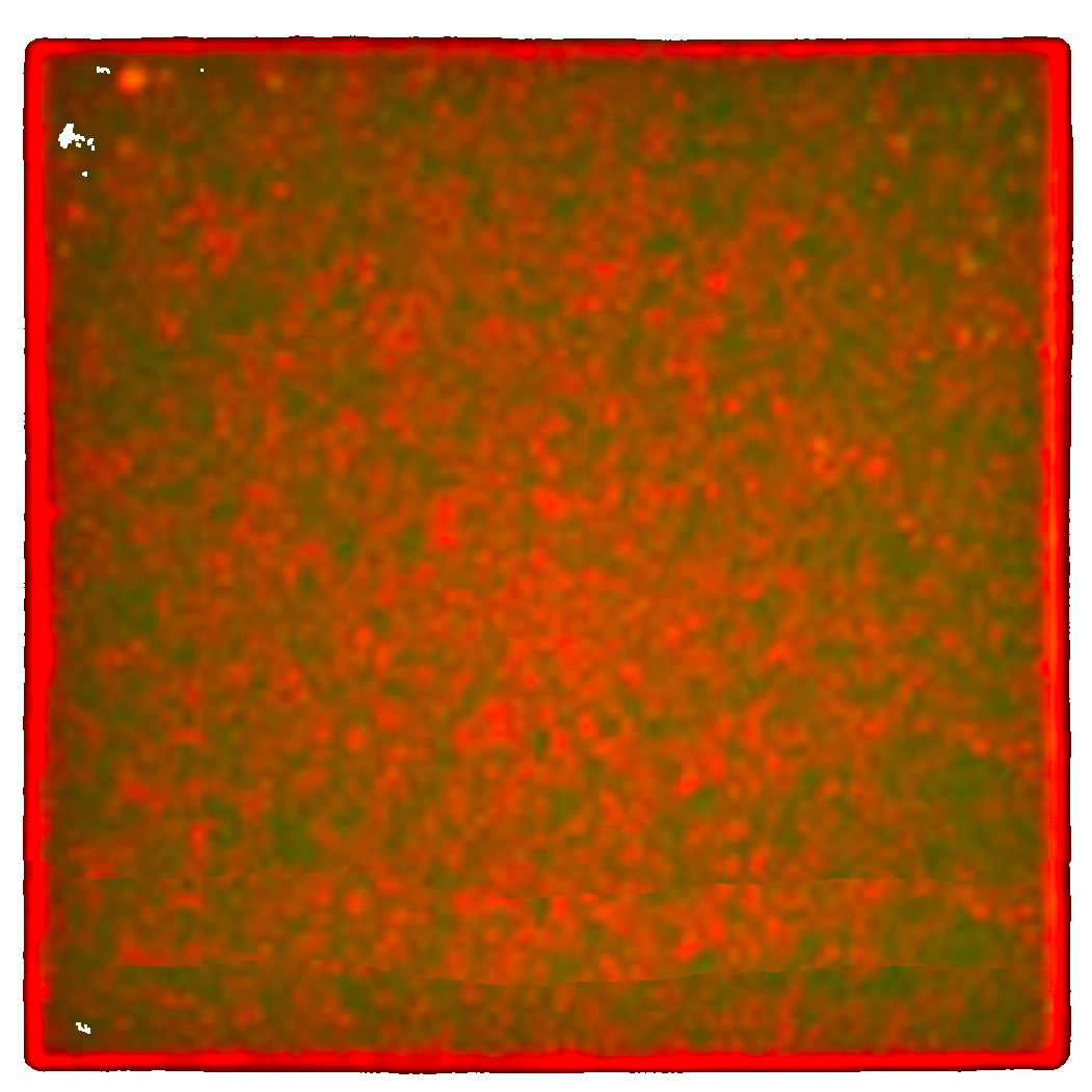
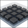
Anti Stokes Thermometry of Plasmonic Nanoparticle Arrays
S. Ezendam, L. Nan, I. L. Violi, S. A. Maier, E. Cortés,* G. Baffou,* J. Gargiulo*
Advanced Optical Materials , 2301496 (2023)
Abstract:Metallic nanoparticles possess strong photothermal responses, especially when illuminated as ensembles due to collective effects. However, accurately quantifying the temperature increase remains a significant challenge, impeding progress in several applications. Anti Stokes thermometry offers a promising solution by enabling direct and non-invasive temperature measurements of the metal without the need for labeling or prior calibration. While Anti Stokes thermometry is successfully applied to individual nanoparticles, its potential to study light-to-heat conversion with plasmonic ensembles remains unexplored. In this study, the theoretical framework and the conditions that must be fulfilled for applying Anti Stokes thermometry to ensembles of nanoparticles are discussed. Then, this technique is implemented to measure the light-induced heating of square arrays of Au nanodisks. The obtained temperature measurements are validated using wavefront microscopy, demonstrating excellent agreement between the two thermometry methods. These results showcase the extension of Anti Stokes thermometry to plasmonic ensembles, highlighting its potential for implementation in the diverse photothermal applications involving these systems.
|
 |
64.-
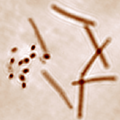
Dry mass photometry of single bacteria using quantitative wavefront microscopy
M. Bénéfice, A. Gorlas, B. Marthy, V. Da Cunha, P. Forterre, A. Sentenac, P. C. Chaumet, G. Baffou*
Biophysical Journal 122, 3159-3172 (2023)
Abstract:Quantitative phase microscopy (QPM) represents a non-invasive alternative to fluorescence microscopy for cell observation with high contrast and for the quantitative measurement of dry mass (DM) and growth rate at the single cell level. While DM measurements using QPM have been widely conducted on mammalian cells, bacteria have been less investigated, presumably due to the high-resolution and high-sensitivity required by their smaller size. This article demonstrates the use of cross-grating wavefront microscopy (CGM), a high-resolution and high-sensitivity QPM, for accurate DM measurement and monitoring of single micro-organisms (bacteria and archaea). The article covers strategies for overcoming light diffraction and sample focusing, and introduces the concepts of normalized optical volume (OV) and optical polarizability (OP) to gain additional information beyond DM. The algorithms for DM, OV, and OP measurements are illustrated through two case studies: monitoring dry mass evolution in a microscale colony forming unit as a function of temperature, and using OP as a potential species-specific signature.
|
 |
63.-
Uniform Huygens Metasurfaces with Postfabrication Phase Pattern
Recording Functionality
E. Mikheeva, R. Colom, P. Genevet*, F. Bedu, I. Ozerov, S. Khadir, G. Baffou, R. Abdeddaim, S. Enoch, and J. Lumeau*
ACS Photonics 10, 1538-1546 (2023)
Abstract:With the rapid progress in the field of metasurfaces and their use in miniature integrated devices arises the quest for cheap mass production of efficient metasurfaces. We suggest a novel way to design and fabricate phase-gradient Huygens metasurfaces using laser-annealing of uniform particles made of As2S3 chalcogenide glass. We show that a phase gradient metasurface can be realized by tuning the refractive index of otherwise identical meta-atoms instead of tuning their geometry. We are using an array of identical As2S3 particles with the possibility to locally change their refractive index using a short-wavelength illumination (green laser) in order to tune the phase pattern at the postfabrication stage. Metasurfaces fabricated with this method can be used for operation in the red or IR spectral range. We fabricate uniform As2S3 Huygens metasurfaces using electron beam lithography and demonstrate their postfabrication tuning with exposure of comparatively low intensity. Sample characterizations with transmittance measurements and quantitative phase microscopy provide results in good correspondence with numerical predictions confirming postfabrication spectral tuning. Using such tuning, we demonstrate the possibility of transferring the intensity pattern produced by modifying a writing beam with a spatial light modulator to a phase pattern recorded on a uniform As2S3 metasurface. Our method has potential advantages for the low-cost production of large-scale metasurfaces because uniform geometries are better adjusted for mass manufacturing.
|
 |
62.-
Wavefront microscopy using quadriwave lateral shearing interferometry: from bioimaging to nanophotonics
G. Baffou
ACS Photonics 10, 322-339 (2023)
Abstract:Common cameras are only sensitive to the intensity of light, discarding an essential feature of a light wave: its phase profile or, equivalently, its wavefront profile. This Review focuses on a rising wavefront imaging technique called quadriwave lateral shearing interferometry (QLSI), based on the simple use of a 2-dimensional diffraction grating, aka a cross-grating, in front of a regular camera. We detail the working principle of QLSI and its implementation on an optical microscope. We highlight its microscopy applications in bioimaging and nanophotonics, in particular for the characterization of living cells, nanoparticles, 2D materials, metasurfaces, microscale temperature gradients, and surface topography. Finally, we draw a critical comparison of QLSI with current quantitative phase microscopy techniques, namely, digital holography microscopy (DHM), spatial light interference microscopy (SLIM), and diffraction phase microscopy (DPM).
|
2022
 |
60.-
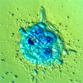
Life at high temperature observed in vitro upon laser heating of gold nanoparticles
C. Molinaro, M. Bénéfice, A. Gorlas, V. Da Cunha, H. M. L. Robert, R. Catchpole, L. Gallais, P. Forterre, G. Baffou*
Nature Communications 13, 5342 (2022)
Abstract: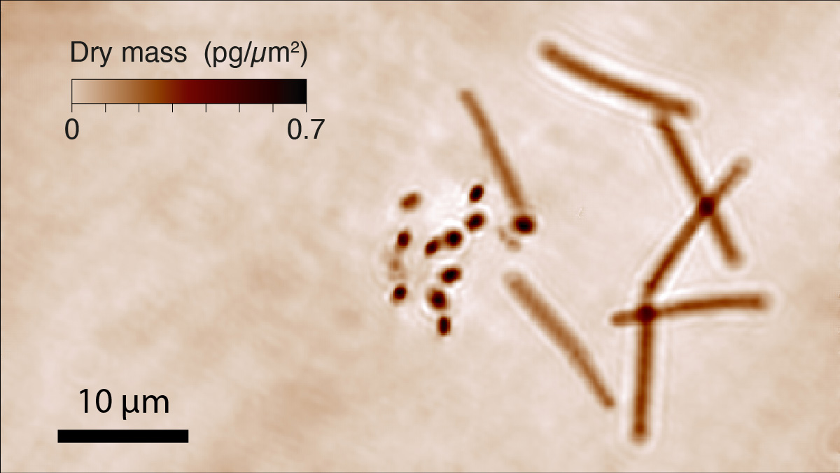
Thermophiles are microorganisms that thrive at high temperature. Studying them can provide valuable information on how life has adapted to extreme conditions. However, high temperature conditions are difficult to achieve on conventional optical microscopes. Some home-made solutions have been proposed, all based on local resistive electric heating, but no simple commercial solution exists. In this article, we introduce the concept of microscale laser heating over the field of view of a microscope to achieve high temperature for the study of thermophiles, while maintaining the user environment in soft conditions. Microscale heating with moderate laser intensities is achieved using a substrate covered with gold nanoparticles, as biocompatible, efficient light absorbers. The influences of possible microscale fluid convection, cell confinement and centrifugal thermophoretic motion are discussed. The method is demonstrated with two species: (i) Geobacillus stearothermophilus, a motile thermophilic bacterium thriving around 65°C, which we observed to germinate, grow and swim upon microscale heating and (ii) Sulfolobus shibatae, a hyperthermophilic archaeon living at the optimal temperature of 80°C. This work opens the path toward simple and safe observation of thermophilic microorganisms using current and accessible microscopy tools.
|
 |
59.-
Cross-grating phase microscopy (CGM): In-silico experiment (insilex) algorithm, noise and accuracy
B. Marthy, G. Baffou*
Optics Communications 521, 128577 (2022)
Abstract:Cross-grating phase microscopy (CGM) is a quantitative phase microscopy technique based on the association of a 2-dimensional diffraction grating (cross-grating) and a regular camera sensor, separated by a millimetric distance. This simple association enables the high-resolution imaging of the complex electric field amplitude of a light beam (intensity and phase) from a single image acquisition. While CGM has been used for metrology applications in cell biology and nanophotonics this last decade, there has been few studies on its basics, especially for the microscopy community. In this article, we provide a numerical algorithm that enables the in silico (i.e. computer-simulated) data acquisition, to easily vary and observe the effects of all the CGM experimental parameters using computer means. In the frame on this article, we illustrate the interest of this numerical algorithm by using it to explain and quantify the effects of several important CGM parameters (grating-camera distance, pixel size, light intensity, numerical apertures, etc.) on the noise, precision and trueness of CGM measurements. This work is aimed to push the limits of CGM toward advanced applications in biomicroscopy and nanophotonics.
|
 |
58.-
Optically-assisted thermophoretic
reversible assembly of colloidal
particles and E. coli using graphene
oxide microstructures
J. Puthenveetil Joby, S. Das, P. Pinapati, B. Rogez,
G. Baffou, D. K. Tiwari, S. Cherukulappurath
Scientific Reports 12, 3657 (2022)
Abstract:Optically?assisted large?scale assembly of nanoparticles have been of recent interest owing to their potential in applications to assemble and manipulate colloidal particles and biological entities. In the recent years, plasmonic heating has been the most popular mechanism to achieve temperature hotspots needed for extended assembly and aggregation. In this work, we present an alternative route to achieving strong thermal gradients that can lead to non?equilibrium transport and assembly of matter. We utilize the excellent photothermal properties of graphene oxide to form a large?scale assembly of silica beads. The formation of the assembly using this scheme is rapid and reversible. Our experiments show that it is possible to aggregate silica beads (average size 385 nm) by illuminating thin graphene oxide microplatelet by a 785 nm laser at low intensities of the order of 50–100 ?W/?m2. We further extend the study to trapping and photoablation of E. coli bacteria using graphene oxide. We attribute this aggregation process to optically driven thermophoretic forces. This scheme of large?scale assembly is promising for the study of assembly of matter under non?equilibrium processes, rapid concentration tool for spectroscopic studies such as surface?enhanced Raman scattering and for biological applications.
|
 |
57.-
Cross-grating phase microscopy for nanophotonics
G. Baffou
arXiv , 2112.14924 (2022)
Abstract:Quantitative phase microscopies (QPMs) have been mainly used for applications in cell biology, for around 2 decades. In this article, we show how cross-grating phase microscopy (CGM), a high-resolution, high-sensitivity QPM, recently expanded the scope of QPMs to applications in nanophotonics. In particular, this article explains how the intensity and phase images acquired by CGM can be processed to determine all the optical properties of imaged nanoparticles, 2D-materials and metasurfaces. We also explain how CGM can be used as a temperature microscopy technique. This latter imaging modality led to a large variety of works in the 2010s based on the optical heating of plasmonic nanoparticles for photothermal studies in physics, chemistry and biology at the microscale, in which label-free, microscale temperature measurements were pivotal.
|
2021
 |
56.-
Thermoplasmonics of metal layers and nanoholes
B. Rogez,* Z. Marmri, F. Thibaudau, G. Baffou*
APL Photonics 6, 101101 (2021)
Abstract:Since the early 2000s, the experimental and theoretical studies of photothermal effects in plasmonics have been mainly oriented toward systems composed of nanoparticles, mostly motivated by applications in biomedecine, and overlooked the case of plasmonic resonances of nanoholes in metal layers (also called nanopores or nano-apertures). Yet, more and more applications based on plasmonic nanoholes have been reported these last years (e.g., optical trapping, molecular sensing, surface-enhanced Raman scattering), and photothermal effects can be unexpectedly high for this kind of systems, mainly because of the very large amount of metal under illumination, compared with nanoparticle systems. Nanoholes in metal layers involve a fully different photothermodynamical picture, and few of what is known about nanoparticles can be applied with nanoholes. A plasmonic nanohole mixes localized and surfaces plasmons, along with heat transport in a two-dimensional highly conductive layer, making the underlying photothermodynamical physics particularly complex. This article is aimed to provide a comprehensive description of the photothermal effects in plasmonics when metal layers are involved, based on experimental, theoretical and numerical results (we share in Suppl. Mater. all the numerical codes used in this article). Photothermal effects in metal layers (embedded or suspended) are first described in detail, followed by the study of nanoholes, where we revisit the concept of absorption cross section, and discuss the influences of parameters such as layer thickness, layer composition, nanohole size and geometry, adhesion layer, thermal radiation, and illumination wavelength.
|
 |
55.-
Microscale Thermophoresis in Liquids Induced by Plasmonic Heating and Characterized by Phase and Fluorescence Microscopies
S. Shakib, B. Rogez, S. Khadir, J. Polleux, A. Würger, G. Baffou*
J Phys Chem C 125, 21533-21542 (2021)
Abstract:Thermophoresis denotes the motion of particles along temperature gradients. Insignificant in most daily life observations, this peculiar effects can become dominant in applications involving nano- and microscale heating in fluids. Recent studies in nanoplasmonics observed significant thermophoresis of molecules and particles, in particular in plasmonic trapping, SERS and biosensing. Evidencing the presence of thermophoresis is not obvious and quantifying its magnitude is even less accessible considering existing techniques. In this article, we introduce a method capable of quantifying the thermophoresis of particles in the context of nanoplasmonic applications. A gold nanoparticle array under illumination is used to create microscale temperature gradients, and a dual fluorescence-phase microscopy technique is used to map both temperature and concentration in parallel. This association enables the determination of Soret coefficients for a wide range of temperature from a single image acquisition. This metrology technique paves the way for broader fundamental research in microscale thermophoresis in liquids, and better controlled applications in nanophotonics involving thermoplasmonic effects.
|
 |
53.-
Quantitative phase microscopy using quadriwave lateral shearing interferometry (QLSI): principle, terminology, algorithm and grating shadow description.
G. Baffou
J. Phys. D: Appl. Phys. 54, 294002 (2021)
Abstract:Quadriwave lateral shearing interferometry (QLSI) is a quantitative phase imaging technique based on the use of a diffraction grating placed in front of a camera. This grating creates a wire-mesh-like image, called an interferogram, that is postprocessed to retrieve both the intensity and phase profiles of an incoming light beam. Invented in the 90s, QLSI has been used in numerous applications, e.g., laser beam characterization, lens metrology, topography measurements, adaptive optics, or gas jet metrology. More recently, the technique has been implemented on optical microscopes to characterize micro and nano-objects for bioimaging and nanophotonics applications. However, not much effort has been placed on disseminating this powerful technology so far, while it is yet a particularly simple technique. In this article, we intend to popularize this technique by describing all its facets in the framework of optical microscopy, namely the working principle, its implementation on a microscope and the theory of image formation, using simple pictures. Also, we provide and comment an algorithm of interferogram processing, written in Matlab. Then, following the new extension of the technique for microscopy and nanophotonics applications, and the deviation from what the technique was initially invented for, we propose to revisit the description of the technique, in particular by discussing the terminology, insisting more on a grating-shadow description rather than a quadriwave process, and proposing an alternative appellation, namely "grating shadow phase microscopy" or "grating-assisted phase microscopy".
|
 |
52.-
Are bacteria claustrophobic? The problem of micrometric spatial confinement for the culture of micro-organisms
C. Molinaro,* V. Da Cunha, A. Gorlas, F. Iv, L. Gallais, R. Catchpole, P. Forterre, G. Baffou*
RSC Advances 11, 12500-12506 (2021)
Abstract:Culturing cells confined in microscale geometries has been reported in many studies this last decade, in particular following the development of microfluidic-based applications and lab-on-a-chip devices. Such studies usually examine growth of Escherichia coli. In this article, we show that E. coli may be a poor model and that spatial confinement can severely prevent the growth of many micro-organisms. By studying different bacteria and confinement geometries, we determine that the growth inhibition observed for some bacteria results from fast dioxygen depletion, inherent to spatial confinement, and not to any depletion of nutriments. This article unravels the physical origin of confinement problems in cell culture, highlighting the importance of oxygen depletion, and paves the way for the effective culture of bacteria in confined geometries by demonstrating enhanced cell growth in confined geometries in the proximity of air bubbles.
|
 |
51.-
Metasurface optical characterization using quadriwave lateral shearing interferometry
S. Khadir,* D. Andrén, R. Verre, Q. Song, S. Monneret, P. Genevet, M. Käll, G. Baffou*
ACS Photonics 8, 603-613 (2021)
Abstract:An optical metasurface consists of a dense and usually non-uniform layer of scattering nanostructures behaving as a continuous and extremely thin optical component, with predefined phase and intensity transmission/reflection profiles. To date, various sorts of metasurfaces (metallic, dielectric, Huygens-like, Pancharatman-Berry, etc.) have been introduced to design ultrathin lenses, beam deflectors, holograms, or polarizing interfaces. Their actual efficiencies depend on the ability to predict their optical properties and to fabricate non-uniform assemblies of billions of nanoscale structures on macroscopic surfaces. To further help improve the design of metasurfaces, precise and versatile post-characterization techniques need to be developed. Today, most of the techniques used to characterize metasurfaces rely on light intensity measurements. Here, we demonstrate how quadriwave lateral shearing interferometry (QLSI), a quantitative phase microscopy technique, can easily achieve full optical characterization of metasurfaces of any kind, as it can probe the local phase imparted by a metasurface with high sensitivity and spatial resolution. As a means to illustrate the versatility of this technique, we present measurements on two types of metasurfaces, namely Pancharatnam-Berry and effective-refractive-index metasurfaces, and present results on uniform metasurfaces, metalenses and deflectors.
|
2020
 |
50.-
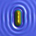
Quantifying the Role of the Surfactant and the Thermophoretic Force in Plasmonic Nano-Optical Trapping
Q. Jiang, B. Rogez, J. B. Claude, G. Baffou, J. Wenger*
Nano Letters 12, 8811-8817 (2020)
Abstract:Plasmonic nanostructures generate intense electric field gradients, enabling efficient optical trapping of nano-objects. However, because part of the incident light is absorbed into the metal, a temperature gradient is present around the plasmonic nano-tweezers, leading to an additional thermophoretic force. As both optical and thermophoretic forces occur simultaneously, investigating their relative contributions remains an intricate challenge for plasmonic trapping. Another issue concerns the role of the surfactant that is added to the solution to avoid aggregation of the nanoparticles. While various surfactants are commonly used in plasmonic trapping, their influence is often ignored. Here we show that the influence of the thermophoretic force and the surfactant role are closely interconnected. The surfactant can significantly impact the trap performance: we measure trap stiffness 20× higher using sodium dodecyl sulfate (SDS) as compared to Triton X-100. This effect stems from the influence of the surfactant in setting the thermophilic or thermophobic response of the nanoparticles, which in turn determines the sign and amplitude of the thermophoretic force. Using different surfactant conditions, we disentangle the thermophoretic contribution from the optical gradient force, and measure the amplitude of each force in nano-optical trapping. This approach can be easily extended to investigate other plasmonic designs. Moreover, it provides clear guidelines to maximize the nano-optical trap performance by properly choosing the surfactant conditions.
|
 |
49.-
Applications and challenges of thermoplasmonics
G. Baffou,* F. Cichos,* R. Quidant*
Nature Materials 19, 946-958 (2020)
Abstract:Over the past two decades, there has been a growing interest in the use of plasmonic nanoparticles as sources of heat remotely controlled by light, giving rise to the field of thermoplasmonics. The ability to release heat on the nanoscale has already impacted a broad range of research activities, from biomedicine to imaging and catalysis. Thermoplasmonics is now entering an important phase: some applications have engaged in an industrial stage, while others, originally full of promise, experience some difficulty in reaching their potential. Meanwhile, innovative fundamental areas of research are being developed. In this Review, we scrutinize the current research landscape in thermoplasmonics, with a specific focus on its applications and main challenges in many different fields of science, including nanomedicine, cell biology, photothermal and hot-electron chemistry, solar light harvesting, soft matter and nanofluidics.
|
 |
48.-
Simple experimental procedures to distinguish photothermal from hot-carrier processes in plasmonics
G. Baffou,* I. Bordacchini, A. Baldi, R. Quidant
Light: Science and Applications 9, 2047-7538 (2020)
☆ESI highly cited paper in January 2023
Abstract:Light absorption and scattering of plasmonic metal nanoparticles can lead to non equilibrium charge carriers, intense electromagnetic near-fields, and heat generation, with promising applications in a vast range of fields, from chemical and physical sensing, to nanomedicine, and photocatalysis for the sustainable production of fuels and chemicals. Disentangling the relative contribution of thermal and non-thermal contributions in plasmon driven processes is however difficult. Nanoscale temperature measurements are technically challenging and macroscale experiments are often characterized by collective heating effects, which tend to make the actual temperature increase unpredictable. This perspective is intended to help the reader experimentally detect and quantify photothermal effects in plasmon-driven chemical reactions, to discriminate their contribution from the one due to photochemical processes, and to cast a critical eye on the current literature. To this aim, we introduce 7 simple experimental procedures, which do not require the use of complex or expensive thermal microscopy techniques. These proposed procedures are adaptable to a wide range of experiments and fields of research where photothermal effects need to be assessed, such as plasmonic-assisted chemistry, heterogeneous catalysis, photovoltaics, biosensing and enhanced molecular spectroscopy.
|
 |
47.-
Optimal architecture for diamond-based wide-field thermal imaging
R. Tanos, W. Akhtar, S. Monneret, F. Favaro de Oliveira, G. Seniutinas, M. Munsch, P. Maletinsky, L. le Gratiet, I. Sagnes, A. Dréau, C. Gergely, V. Jacques, G. Baffou, I. Robert-Philip
AIP Advances 10, 025027 (2020)
Abstract:Nitrogen-Vacancy centers in diamond possess an electronic spin resonance that strongly depends on temperature, which makes them efficient temperature sensor with a sensitivity down to a few mK/√Hz. However, the high thermal conductivity of the host diamond may strongly damp any temperature variations, leading to invasive measurements when probing local temperature distributions. In view of determining possible and optimal configurations for diamond-based wide-field thermal imaging, we here investigate, both experimentally and numerically, the effect of the presence of diamond on microscale temperature distributions. Three geometrical configurations are studied: a bulk diamond substrate, a thin diamond layer bonded on quartz and diamond nanoparticles dispersed on quartz. We show that the use of bulk diamond substrates for thermal imaging is highly invasive, in the sense that it prevents any substantial temperature increase. Conversely, thin diamond layers partly solve this issue and could provide a possible alternative for microscale thermal imaging. Dispersions of diamond nanoparticles throughout the sample appear as the most relevant approach as they do not affect the temperature distribution, although NV centers in nanodiamonds yield lower temperature sensitivities compared to bulk diamond.
|
 |
45.-
Adhesion Layer Influence on Controlling the Local Temperature in Plasmonic Gold Nanoholes
Q. Jiang, B. Rogez, J.-B. Claude, A. Moreau, J. Lumeau, G. Baffou, J. Wenger*
Nanoscale 12, 2524-2531 (2020)
Abstract:Gold films do not adhere well on glass substrates, so plasmonics experiments typically use a thin adhesion layer of titanium or chromium to ensure a proper adhesion between the gold film and the glass substrate. While the absorption of light into gold structures is largely used to generate heat and control the temp- erature at the nanoscale, the influence of the adhesion layer on this process is largely overlooked. Here, we quantify the role of the adhesion layer in determining the local temperature increase around a single nanohole illuminated by a focused infrared laser. Despite their nanometer thickness, adhesion layers can absorb a greater fraction of the incoming infrared light than the 100 nm thick gold layer leading to a sig- nificant increase of the local temperature. Different experimental designs are explored, offering new ways to promote or avoid the temperature increase inside nanoapertures. This knowledge further expands the plasmonic toolbox for temperature-controlled experiments including single molecule sensing, nanopore translocation, polymerization, or nano-optical trapping.
|
2019
 |
44.-
Temperature Measurement in Plasmonic Nanoapertures used for Optical Trapping
Q. Jiang, B. Rogez, J.-B. Claude, G. Baffou, J. Wenger*
ACS Photonics 6, 1763-1773 (2019)
Abstract:Plasmonic nanoapertures generate strong field gradients enabling efficient optical trapping of nano-objects. However, because the infrared laser used for trapping is also partly absorbed into the metal leading to Joule heating, plasmonic nano-optical tweezers face the issue of local temperature increase. Here, we develop three independent methods based on molecular fluorescence to quantify the temperature increase induced by a 1064 nm trapping beam focused on single and double nanoholes milled in gold films. We show that the temperature in the nanohole can be increased by 10°C even at the moderate intensities of 2 mW/µm 2 used for nano-optical trapping. The temperature gain is found to be largely governed by the Ohmic losses into the metal, independently of the aperture size, double-nanohole gap or laser polarization. The techniques developed therein can be readily extended to other structures to improve our understanding of nano-optical tweezers and explore heat-controlled chemical reactions in nanoapertures.
|
 |
43.-
Microscale Temperature Shaping Using Spatial Light Modulation on Gold Nanoparticles
L. Durdevic, H. M. L. Robert, B. Wattellier, S. Monneret, G. Baffou*
Scientific Report 9, 4644 (2019)
Abstract:Heating on the microscale using focused lasers gave rise to recent applications, e.g., in biomedicine, biology and microfluidics, especially using gold nanoparticles as efficient nanoabsorbers of light. However, such an approach naturally leads to nonuniform, Gaussian-like temperature distributions due to the diffusive nature of heat. Here, we report on an experimental means to generate arbitrary distributions of temperature profiles on the micrometric scale (e.g. uniform, linear, parabolic, etc) consisting in illuminating a uniform gold nanoparticle distribution on a planar substrate using spatially contrasted laser beams, shaped using a spatial light modulator (SLM). We explain how to compute the light pattern and the SLM interferogram to achieve the desired temperature distribution, and demonstrate the approach by carrying out temperature measurements using quantitative wavefront sensing.
|
2018
 |
41.-
Photothermal control of heat-shock protein expression at the single cell level
H. M. L. Robert,* J. Savatier, S. Vial, J. Verghese, B. Wattelier, H. Rigneault, S. Monneret, J. Polleux,* and G. Baffou*
Small 14, 1801910 (2018)
Abstract:Laser heating of individual cells in culture recently led to seminal studies in cell poration, fusion, migration or nanosurgery, although measuring the local temperature increase in such experiments remains a challenge. Here, we demonstrate the laser-induced dynamical control of the heat-shock response at the single cell level, enabled by the use of light-absorbing gold nanoparticles as nanosources of heat and a temperature mapping technique based on quadriwave lateral shearing interferometry (QLSI) measurements. As it is label-free, this approach does not suffer from artifacts inherent to previously reported fluorescence-based temperature-mapping techniques and enables the use of any standard fluorescent labels to monitor in parallel the cell's response.
|
2017
 |
39.-
Optical imaging and characterization of graphene and other 2D materials using quantitative phase microscopy
S. Khadir,* P. Bon, D. Vignaud, E. Galopin, N. McEvoy, D. McCloskey, S. Monneret, G. Baffou*
ACS Photonics 4, 3130-3139 (2017)
Abstract:This article introduces an optical microscopy technique for the characterization of two-dimensional (2D) materials. The technique is based on the use of quadriwave lateral shearing interferometry (QLSI), a quantitative phase imaging technique that allows the imaging of both the intensity and the phase of an incoming light beam. The article shows how QLSI can be used to (i) image 2D materials with high contrast on transparent substrates, (ii) detect the presence of residues coming from the fabrication process and (iii) map the 2D complex optical conductivity and complex refractive index by processing the intensity and phase images of a light beam crossing the 2D material of interest. To illustrate the versatility of this approach for 2D material imaging and characterization, measurements have been performed on graphene and MoS2.
|
2016
 |
36.-
Plasmonic efficiencies of nanoparticles made of metal nitrides (TiN, ZrN) compared with gold
A. Lalisse, G. Tessier, J. Plain, G. Baffou*
Scientific Reports 6, 38647 (2016)
Abstract:Metal nitrides have been proposed to replace noble metals in plasmonics for some specific applications. In particular, while titanium nitride (TiN) and zirconium nitride (ZrN) possess localized plasmon resonances very similar to gold in magnitude and wavelength, they benefit from a much higher sustainability to temperature. For this reason, they are foreseen as ideal candidates for applications in nanoplasmonics that require high material temperature under operation, such as heat assisted magnetic recording (HAMR) or thermophotovoltaics. This article presents a detailed investigation of the plasmonic properties of TiN and ZrN nanoparticles in comparison with gold nanoparticles, as a function of the nanoparticle morphology. As a main result, metal nitrides are shown to be poor near-field enhancers compared to gold, no matter the nanoparticle morphology and wavelength. The best efficiencies of metal nitrides as compared to gold in term of near-field enhancement are obtained for small and spherical nanoparticles, and they do not exceed 60%. Nanoparticle enlargements or asymmetries are detrimental. These results mitigate the utility of metal nitrides for high-temperature applications such as HAMR, despite their high temperature sustainability. Nevertheless, at resonance, metal nitrides behave as efficient nanosources of heat and could be relevant for applications in thermoplasmonics, where heat generation is not detrimental but desired.
|
 |
35.-
Light-Assisted Solvothermal Chemistry Using Plasmonic Nanoparticles
H. M. L. Robert,* F. Kundrat, E. Bermudez-Urena, H. Rigneault, S. Monneret, R. Quidant, J. Polleux, G. Baffou*
ACS Omega 1, 2-8 (2016)
☆ Highlighted on the CNRS website
Abstract: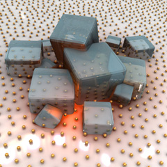
Solvothermal synthesis, denoting chemical reactions occurring in metastable liquids above their boiling point, normally requires the use of a sealed autoclave under pressure, to prevent the solvent from boiling. This work introduces an experimental approach that enables solvothermal synthesis at ambient pressure, in an open reaction medium. The approach is based on the use of gold nanoparticles deposited on a glass substrate and acting as photothermal sources. To illustrate the approach, the selected hydrothermal reaction involves the formation of indium hydroxide microcrystals favored at 200°C in liquid water. In addition to demonstrating the principle, the benefits and the specific characteristics of such an approach are investigated, in particular the much faster reaction rate, the achievable spatial and time scales, the effect of microscale temperature gradients, the effect of the size of the heated area and the effect of thermal-induced microscale fluid convection. This technique is general and could be used to spatially control the deposition of virtually any material for which a solvothermal synthesis exists.
|
2015
 |
33.-
Quantifying the Efficiency of Plasmonic Materials for Near-Field Enhancement and Photothermal Conversion
A. Lalisse, G. Tessier, J. Plain, G. Baffou*
Journal of Physical Chemistry C 119, 25518-25528 (2015)
Abstract:Following recent advances in nanoplasmonics related to high-temperature applications, hot-electron processes, nanochemistry, sensing and active plasmonics, new materials have been introduced, reducing the supremacy of gold and silver in plasmonics. The variety of possible materials in nanoplasmonics is now so wide that selecting the best material for a specific application at a specific wavelength may become a difficult task. In this context, we introduce in this article two dimensionless parameters acting as figures of merit to simply compare the plasmonic capabilities of different materials. These numbers, which we named Faraday and Joule numbers, aim at quantifying the ability of a nanoparticle to respectively enhance the optical near field and produce heat. The benefit of these numbers compared to previously defined figures of merit is that (i) they possess simple close-form expressions and can be simply calculated without numerical simulations (ii) they give quantitative estimations in the non-retarded regime and (iii) they take into account the nature of the surrounding medium. Within this article, we address a wide variety of materials, namely gold, silver, aluminum, copper, cobalt, chromium, iron, molybdenum, manganese, nickel, palladium, platinum, rhodium, tantalum, titanium, titanium nitride, tungsten and zirconium nitride.
|
2014
 |
28.-
Time-harmonic optical heating of plasmonic nanoparticles
P. Berto, M. S. A. Mohamed, H. Rigneault, G. Baffou*
Physical Review B 90, 035439 (2014)
Abstract:Under illumination at their plasmonic resonance wavelength, metal nanoparticles can turn into efficient nanosources of heat by light absorption. Heating a small volume makes it possible to achieve fast dynamics. In this article, we investigate theoretically, numerically and experimentally the temperature distribution of a plasmonic system generated by a modulated incoming light. In particular, we study the response in amplitude and phase of the temperature variations. The cases of single and multiple nanoparticles are both addressed. Many parameters are discussed such as the nature of the media (nanoparticle and surroundings), the size of the nanoparticle or of the plasmonic system, the nanoparticle interdistance, the frequency of the modulation, a possible finite surface thermal conductivity of the nanoparticles and the dimensionality of the system. This work is also intended to determine how fast a plasmonic system is able to induce temperature variations in its surrounding medium.
|
2013
 |
23.-
Three-dimensional temperature imaging around a gold microwire
P. Bon, N. Belaid, D. Lagrange, C. Bergaud, H. Rigneault, S. Monneret, G. Baffou*
Applied Physics Letters 102, 244103 (2013)
Abstract:We report on the temperature mapping around a resistively heated gold microwire. The temperature is determined by measuring the thermal-induced distortion of an incident optical wavefront crossing the system. The optical technique we introduce herein allows, in addition to 3-dimensional temperature measurements, a retrieval of the heat source density at optical resolution. Experimental results are supported by finite element simulations and electric measurements. Applications are envisioned in microelectronics, microfluidics or nanochemistry.
|
 |
22.-
Thermo-plasmonics: using metallic nanostructures as nano-sources of heat
G. Baffou,* R. Quidant*
Laser and Photonics Reviews 7, 171-187 (2013)
☆ Highlighted in Laser & Photon. Rev. as the 3rd most cited article published in 2013
Abstract:
Recent years have seen a growing interest in using metal nanostructures to control temperature on the nanoscale. Under illumination at its plasmonic resonance, a metal nanoparticle features enhanced light absorption turning it into an ideal nano-source of heat, remotely controllable by light. Such a powerful and flexible photothermal scheme is the basis of Thermo-plasmonics. Here, the recent progress of this emerging and fast-growing field is reviewed. First, the physics of heat generation in metal nanoparticles is described, both under continuous or pulsed illumination. A second part is dedicated to numerical and experimental methods that have been developed to further understand and engineer plasmonic-assisted heating processes on the nanoscale. Finally, some of the most recent applications based on the heat generated by gold nanoparticles are surveyed, namely photothermal cancer therapy, nano-surgery, drug delivery, photothermal imaging, protein tracking, photoacoustic imaging, nano-chemistry and optofluidics.
|
2012
 |
18.-
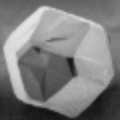
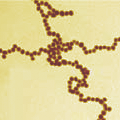
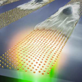
Plasmonic Nanoparticle Networks for Light and Heat Concentration
A. Sanchot, G. Baffou, R. Marty, A. Arbouet, R. Quidant*, C. Girard, E. Dujardin*
ACS Nano 6, 3434-3440 (2012)
Abstract:Self-assembled Plasmonic Nanoparticle Networks (PNN) composed of chains of 12-nm diameter crystalline gold nanoparticles exhibit a longitudinally coupled plasmon mode centered at 700 nm. We have exploited this longitudinal absorption band to efficiently confine light fields and concentrate heat sources in the close vicinity of these plasmonic chain networks. The mapping of the two phenomena on the same superstructures was performed by combining two-photon luminescence (TPL) and fluorescence polarization anisotropy (FPA) in light-induced hyperthermia applications. Furthermore, we propose a unified theoretical process driven by interparticle dipolar interactions.3,5,6 In suspension, these superstructures exhibit an overall globular size of typically 2-3 ?m but individual chain segments are one-nanoparticle, i.e. 12 nm, wide. As illustrated in Figure 1, the optical absorption spectra of these superstructures, that we call Plasmonic Nanoparticle Networks (PNN), display not only a transverse plasmon mode imaging techniques. Besides the light and heat concentration, we show experimentally that the planar spatial distribution of optical field intensity can be simply modulated by controlling the linear polarization of the incident optical excitation. On the contrary, the heat production, which is obtained here by exciting the structures within the optically transparent window of biological tissues, is evenly spread over the entire PNN. This contrasts with the usual case of localized heating in continuous nanowires, thus opening opportunities for these networks
framework to account for both the non-linear optical and thermal near-fields around PNN. The associated numerical simulations, based on a Green s function formalism, are in excellent
agreement with the experimental images. This formalism therefore provides a versatile tool for the accurate engineering of optical and thermodynamical properties of complex plasmonic colloidal architectures.
|
2011
2010
 |
11.-
Nanoscale control of optical heating in complex plasmonic systems
G. Baffou, R. Quidant, F. J. García de Abajo*
ACS Nano 4, 709 (2010)
☆ Selected for the Virtual Issue on Plasmonics in ACS Nano
Abstract: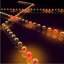
We introduce a numerical technique to investigate the temperature distribution in arbitrarily
complex plasmonic systems subject to external illumination. We perform both electromagnetic
and thermodynamic calculations based upon a time-effcient boundary element method. Two
kinds of plasmonic systems are investigated in order to illustrate the potential of such a technique.
First, we focus on individual particles with various morphologies. In analogy with electrostatics, we
introduce the concept of thermal capacitance. This geometry-dependent quantity allows us to assess
the temperature increase inside a plasmonic particle from the solely knowledge of its absorption
cross section. We present universal thermal-capacitance curves for ellipsoids, rods, disks, and rings.
Additionally, we investigate assemblies of nanoparticles in close proximity and show that, despite
its diffusive nature, the temperature distribution can be made highly non-uniform even at the
nanoscale using plasmonic systems. A significant degree of nanoscale control over the individual
temperatures of neighboring particles is demonstrated, depending on the external light wavelength
and direction of incidence. We illustrate this concept with simulations of gold sphere dimers and
chains in water. Our work opens new possibilities for selectively controlling processes such as local
melting for dynamic patterning of textured materials, chemical and metabolic thermal activation,
and heat delivery for producing mechanical motion with spatial precision in the nanoscale.
|
2009
2008
 |
7.-
Shaping and manipulation of light fields with bottom-up plasmonic structures
C. Girard,* E. Dujardin, G. Baffou, R. Quidant
New Journal of Physics 10, 105016 (2008)
Abstract: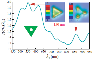
A new interdisciplinary topic which aims at self-assembling, interconnecting and characterizing resonant metallic nanostructures able to funnel, confine, and propagate light energy from a conventional laser source to a single molecular entity is currently emerging in different laboratories. With this technique, several orders of magnitude in the miniaturization scale of optical devices, spanning from tens of micrometres down to the molecular scale, can be expected. With the main objective of overcoming the current limitations of an exclusive top-down approach to plasmonics, we present in this paper some recent experimental and theoretical results about plasmonic structures made by self-assembling or surface deposition of colloidal metallic particles. More specifically, the interest of these objects for tailoring original near-field optical properties will be exposed (near-field optical confinement, local density
of electromagnetic state squeezing, etc). In particular, it is shown that a bottomup approach is not only able to produce interesting nanoscale building blocks but also able to easily produce complex superstructures that would be difficult to achieve by other means.
|
2007
2005
 |
2.-
Scanning tunnelling microscopy imaging and spectroscopy of p-type degenerate 4H-SiC(0001)
A. Laikhtman, G. Baffou, A.J. Mayne, G. Dujardin*
Journal of Physics: Condensed Matter 17, 4015 (2005)
Abstract:
In this work we present scanning tunnelling microscopy (STM) imaging and spectroscopy of a highly p-doped wide bandgap semiconducting 4H-SiC(0001) surface. Whereas n- and p-doped 6H-SiC or n-doped 4H-SiC surfaces can be relatively easily imaged with the STM, the p-doped 4H-SiC cannot be imaged due to the absence of any surface conductivity. This is very surprising given the presence of a p-doped, degenerate epitaxial layer. The behaviour can be explained by the formation of a Schottky barrier either between the tip and the surface or between the surface and the sample holder, depending on the polarity of the applied voltage. We found that prolonged and repeated exposures of the SiC surface to a Si atomic flux followed by thermal annealing are required before the surface conductivity is sufficient to allow STM images to be recorded. The result is the deposition of overlayers of Si, with structures similar to Si(111)7×7, Si(113)3×2, and Si(110)16×2 rather than the expected stable SiC(0001)3×3 reconstruction. We have further demonstrated the ability of scanning tunnelling spectroscopy to distinguish between the Si and the SiC phases based on the difference in their bandgaps.
|
2004
 |
1.-
Chemisorbed bistable molecule: Biphenyl on Si(100)2x1
A.J. Mayne, M.Lastapis, G. Baffou, L. Soukiassian, G. Comtet, L. Hellner, G. Dujardin*
Physical Review B 69, 045409 (2004)
Abstract:
We have shown that the room-temperature adsorption of the biphenyl molecule on the Si(100) surface gives rise in majority to a bistable molecular configuration. The switching of the bistable molecule is activated at room temperature by thermal activation. By using a combination of room-temperature and low-temperature (30 K) scanning tunneling microscope (STM) topography, room-temperature STM manipulation, and near edge x-ray absorption fine structure spectroscopy, the nature of the bistable molecule, its adsorption geometry, and its interaction with the surface could be identified.
|
Fatal error: Uncaught Error: Call to undefined function mysql_close() in /var/www/clients/client1/web2/web/publications.php:339
Stack trace:
#0 {main}
thrown in
/var/www/clients/client1/web2/web/publications.php on line
339


 This article introduces a procedure aimed to quantitatively measure the optical properties of nanoparticles, namely the complex polarizability and the extinction, scattering and absorption cross sections, simultaneously. The method is based on the processing of intensity and wavefront images (PIWI) of a light beam illuminating the nanoparticle of interest. Intensity and wavefront measurements are carried out using quadriwave lateral shearing interferometry, a quantitative phase imaging technique with high spatial resolution and sensitivity. The method does not require any pre-knowledge on the particle, and involves a single interferogram image acquisition. The full determination of the actual optical properties of nanoparticles is of particular interest in plasmonics and nanophotonics for the active search and characterization of new materials, e.g., aimed to replace noble metals in future applications of nanoplasmonics with less-lossy or refractory materials.
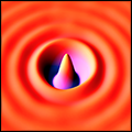
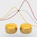
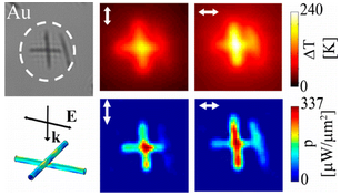
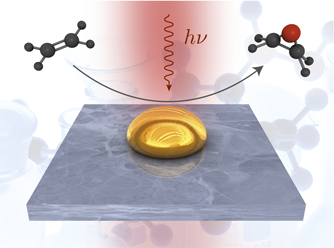
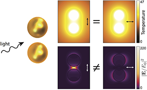 This article introduces the concept of photothermal isosbesticity in plasmonics. In analogy with absorbance spectroscopy, this concept designates nanostructures that feature an invariance of their temperature increase upon varying the illumination polarization angle. We show that non-trivial i.e. non-centrosymmetric) isosbestic nanostructures exist, and prove valuable when the optical near-field intensity remains, on the contrary, highly dependent on the illumination polarization. The concept is introduced with the case of a sphere-dimer, where the conditions for isosbesticity can be derived analytically. The cases of a spheroid and a disc-dimer are also studied in order to draw a general theory and explain how isosbesticity conditions can be obtained from the visible to the infrared range. Non-trivial isosbestic plasmonic nanostructures stand for powerful systems to elucidate the origin (thermal or optical) of mechanisms involved in plasmonics-assisted nanochemistry, liquid-gas phase transition or heat-assisted magnetic recording.
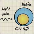
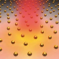
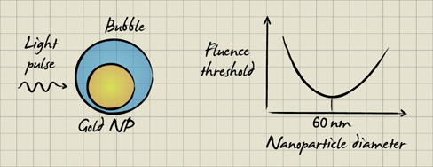 Under nano- to femtosecond pulsed illumination at their plasmonic resonance wavelength, metal nanoparticles efficiently absorb the incident light energy that is subsequently converted into heat. In a liquid environment, with sufficiently high pulse fluences (light energy per unit area), this heat generation may result in the local formation of a transient nanobubble. This phenomenon has been the subject of a decade of investigations and is at the basis of numerous applications from cancer therapy to photoacoutic imaging. The aim of this article is to clarify the question of the fluence threshold required for bubble formation. Using a Runge-Kutta-4 numerical algorithm modeling the heat diffusion around a spherical gold nanoparticle, we numerically investigate the influence of the nanoparticle diameter, pulse duration (from the femto- to the nanosecond range), wavelength and Kapitza resistivity in order to explain the observations reported in the literature.
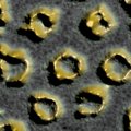
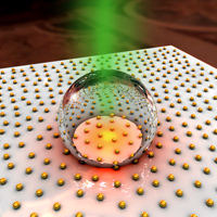
 Shaping and positioning noble metal nanostructures are essential processes that still require laborious and
sophisticated techniques to fabricate functional plasmonic interfaces. The present study reports a simple
photochemical approach compatible with micellar nanolithography and photolithography that enables
the growth, arrangement and shaping of gold nanoparticles with tuneable plasmonic resonances on glass
substrates. Ultraviolet illumination of surfaces coated with gold-loaded micelles leads to the formation of
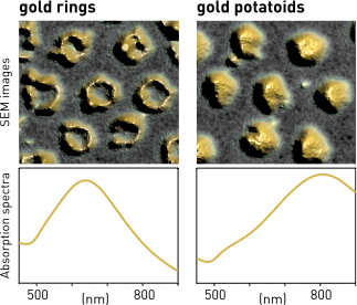
gold nanoparticles with micro/nanometric spatial resolution without requiring any photosensitizers or photoresists. Depending on the extra-micellar chemical environment and the illumination wavelength, block copolymer micelles act as reactive and light-responsive templates, which enable to grow gold deformed nanoparticles (potatoids) and nanorings. Optical characterization reveals that arrays of indivi-
dual potatoids and rings feature a localized plasmon resonance around 600 and 800 nm, respectively,
enhanced photothermal properties and high temperature sustainability, making them ideal platforms for
future developments in nanochemistry and biomolecular manipulation controlled by near-infrared-
induced heat.
Shaping and positioning noble metal nanostructures are essential processes that still require laborious and
sophisticated techniques to fabricate functional plasmonic interfaces. The present study reports a simple
photochemical approach compatible with micellar nanolithography and photolithography that enables
the growth, arrangement and shaping of gold nanoparticles with tuneable plasmonic resonances on glass
substrates. Ultraviolet illumination of surfaces coated with gold-loaded micelles leads to the formation of
gold nanoparticles with micro/nanometric spatial resolution without requiring any photosensitizers or photoresists. Depending on the extra-micellar chemical environment and the illumination wavelength, block copolymer micelles act as reactive and light-responsive templates, which enable to grow gold deformed nanoparticles (potatoids) and nanorings. Optical characterization reveals that arrays of indivi-
dual potatoids and rings feature a localized plasmon resonance around 600 and 800 nm, respectively,
enhanced photothermal properties and high temperature sustainability, making them ideal platforms for
future developments in nanochemistry and biomolecular manipulation controlled by near-infrared-
induced heat.
 In this article, we present a comprehensive investigation of the photothermal properties of plasmonic nanowire networks. We measure the local steady state temperature increase, heat source density and absorption in Ag, Au and Ni metallic nanowire networks under optical illumination. This allows direct experimental confirmation of increased heat generation at the junction between two metallic nanowires, and stacking dependent absorption of polarized light. Due to co-operative thermal effects, the local temperature distribution in a network is shown to be completely delocalized on a micrometer scale, despite the nanoscale features in the heat source density. The steady state temperature rise is shown to scale linearly with the illumination diameter, allowing calibration of the local temperature field. The total illumination area is thus identified as an important parameter controlling local temperature rise, often not considered in thermoplasmonic experiments. Comparison of the experimental temperature profile with numerical simulation allows an upper limit for the effective thermal conductivity of an Ag nanowire network to be established at 43 Wm-1K-1 (0.1 bulk).
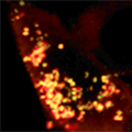
 We introduce a deterministic procedure, named TSUNA for Temperature Shaping Using Nanoparticle Assemblies, aimed at generating arbitrary temperature distributions on the microscale. The strategy consists in (i) using an inversion algorithm to determine the exact heat source density necessary to create a desired temperature distribution and (ii) reproducing experimentally this calculated heat source density using smart assemblies of lithographic metal nanoparticles under illumination at their plasmonic resonance wavelength. The feasibility of this approach was demonstrated experimentally by thermal microscopy based on wavefront sensing.
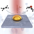
 Noble metal nanoparticles supporting plasmonic resonances behave as efficient nanosources of light, heat and energetic electrons. Owing to these properties, they offer a unique playground to trigger chemical reactions on the nanoscale. In this tutorial review, we discuss how nanoplasmonics can benefit chemistry and review the most recent developments along this new and fast growing field of research.
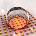
 Under illumination, metal nanoparticles can turn into ideal nanosources of heat due to enhanced light absorption at the plasmonic resonance wavelength. In this article, we aim at providing a comprehensive description of the generation of micro-bubbles in a liquid occurring around plasmonic nanoparticles under continuous illumination. We focus on a common situation where the nanoparticles are located on a solid substrate and immersed in water. Experimentally, we evidenced a series of singular phenomena: (i) the bubble life-time after heating can reach several minutes, (ii) the bubbles are not made of water steam but of air and (iii) the local temperature required to trigger bubble generation is much larger than 100°C: This last observation evidences that superheated liquid water, up to 220°C, is easy to achieve in plasmonics, under ambient pressure conditions and even over arbitrary large areas. This could lead to new chemical synthesis approaches in solvothermal chemistry.
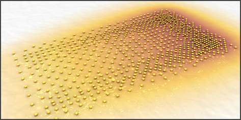
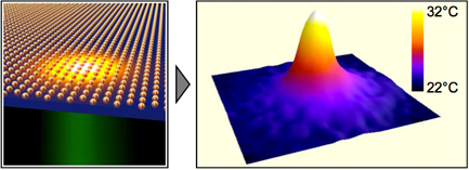
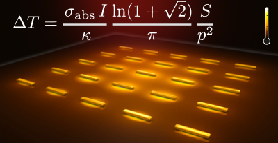 The temperature distribution throughout arrays of illuminated metal nanoparticles is investigated numerically and experimentally. The two cases of continuous and fs-pulsed illumination are addressed. In the case of continuous illumination, two distinct regimes are evidenced: a temperature confinement regime - where the temperature increase remains confined at the vicinity of each nanosource of heat - and a temperature delocalization regime - where the temperature is uniform throughout the whole nanoparticle assembly despite of their nanometric size. We show that the occurrence of one regime or another simply depends on the geometry of the nanoparticle distribution. In particular, we derived simple expressions of i) dimensionless parameters aimed at predicting the degree of temperature confinement and ii) analytical expressions aimed at estimating the actual temperature increase at the centre of an assembly of nanoparticles under illumination, preventing heavy numerical simulations. All these theoretical results are supported by experimental measurements of the temperature distribution on regular arrays of gold nanoparticles under illumination. In the case of fs-pulsed illumination, we explain what are the two conditions that must be fulfilled to observe a further enhanced temperature spatial confinement.
 The ability to reversibly control the interactions between the extracellular matrix (ECM) and cell surface receptors such as integrins would allow one to investigate reciprocal signaling circuits between cells and their surrounding environment. Engineering microstructured culture substrates functionalized with switchable molecules remains the most adopted strategy to manipulate surface adhesive properties, although these systems exhibit limited reversibility and require sophisticated preparation procedures. Here, we report a straightforward protocol to fabricate biofunctionalized micropatterned gold nanoarrays that favor one-dimensional cell migration and function as plasmonic nanostoves to physically block and orient the formation of new adhesion sites. Being reversible and not restricted spatiotemporally, thermoplasmonic approaches will open new opportunities to further study the complex connections between ECM and cells.
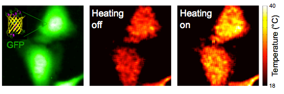 Heat is of fundamental importance in many cellular processes such as cell metabolism, cell division and gene expression. Accurate and non-invasive monitoring of temperature changes in individual cells could thus help clarify intricate cellular processes and develop new applications in biology and medicine. Here we report the use of green fluorescent proteins (GFPs) as a thermal nanoprobes suited for intracellular temperature mapping. Temperature probing is achieved by monitoring the fluorescence polarization anisotropy of GFP. The method is tested on GFP-transfected HeLa and U-87 MG cancer cell lines where we monitored the heat delivery by photothermal heating of gold nanorods surrounding the cells. A spatial resolution of 300 nm and a temperature accuracy of about 0.4°C are achieved. Benefiting from its full compatibility with widely used GFP-transfected cells, this approach provides a non-invasive tool for fundamental and applied research in areas ranging from molecular biology to therapeutic and diagnostic studies.
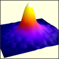
 We introduce an optical microscopy technique aiming at characterizing the heat generation arising from nanostructures, in a comprehensive and quantitative manner. Namely, it permits to i) map the temperature distribution around the source of heat, ii) map the heat power density delivered by the source and iii) retrieve its absorption cross section in the case of a light-absorbing structure. The technique is based on the measure of the thermal-induced refractive index variation of the medium surrounding the source of heat. The measurement is achieved using an association of a regular CCD camera along with a modified Hartmann diffraction grating. Such a simple association makes this technique straightforward to implement on any conventional microscope with its native broadband illumination conditions. We exemplify this technique on gold nanoparticles illuminated at their plasmonic resonance. The spatial resolution of this technique is diffraction limited and temperature variations weaker than 1 K can be detected.
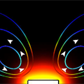
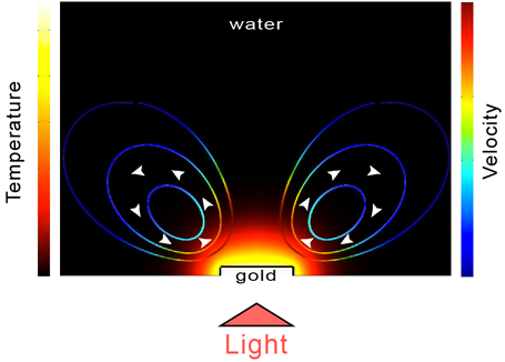 We study the ability of a plasmonic structure under illumination to release heat and induce fluid convection at the nanoscale. We first introduce the unified formalism associated to this multidisciplinary problem combining optics, thermodynamics and hydrodynamics. On this basis, numerical simulations were performed to compute the temperature field and velocity field evolutions of the surrounding fluid for a gold disk on glass while illuminated at its plasmon resonance. We show that the velocity amplitude of the surrounding fluid has a linear dependence on the structure temperature and a quadratic dependence on the structure size (for a given temperature). The fluid velocity remains negligible for single nanometer-sized plasmonic structures (<1 nm/s) due to very low Reynolds number. However thermal-induced fluid convection can play a significant role when considering either micrometer-size structures or an assembly of nanostructures.
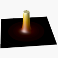
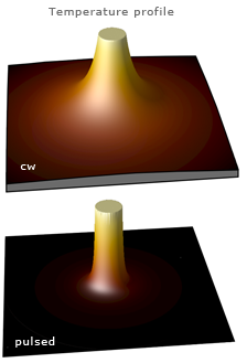 We investigate theoretically and numerically the thermodynamics of gold nanoparticles immersed in water and illuminated by a femtosecond-pulsed laser at their plasmonic resonance. The spatio-temporal evolution of the temperature profile inside and outside is computed using a numerical framework based on a Runge-Kutta algorithm of the fourth order. The aim is to provide a comprehensive description of the physics of heat release of plasmonic nanoparticles under pulsed illumination, along with a simple and powerful numerical algorithm. In particular, we investigate the amplitude of the initial instantaneous temperature increase, the physical differences between pulsed and cw illuminations, the time scales governing the heat release into the surroundings, the spatial extension of the temperature distribution in the surrounding medium, the influence of a finite thermal conductivity of the gold/water interface, the influence of the pulse repetition rate of the laser, the validity of the uniform temperature approximation in the metal nanoparticle, and the optimum nanoparticle size (around 40nm) to achieve maximum temperature increase.
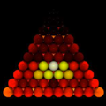
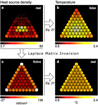 We extend the Discrete Dipole Approximation (DDA) and the Green Dyadic Tensor (GDT) methods - previously dedicated to all-optical simulations - to investigate the thermodynamics of illuminated plasmonic nanostructures. This extension is based on the use of the thermal Green function and a new algorithm that we named Laplace Matrix Inversion. It allows for the computation of the steady-state temperature distribution throughout plasmonic systems. This hybrid photo-thermal numerical method is suited to investigate arbitrarily complex structures, can take into account the presence of a dielectric planar substrate and is simple to implement in any DDA or GDT code. Using this new numerical framework, different applications are discussed such as thermal collective effects in nanoparticles assembly, the influence of a substrate on the temperature distribution and the heat generation in a plasmonic nano-antenna. This numerical approach appears particularly suited for new applications in physics, chemistry and biology such as plasmon-induced nano-chemistry and catalysis, nano-fluidics, photo-thermal cancer therapy or phase-transition control at the nanoscale.
 We investigate the physics of photo-induced heat generation in plasmonic structures by using a novel thermal microscopy technique based on molecular fluorescence polarization anisotropy. This technique enables to image the heat source distribution in light-absorbing systems like plasmonic nanostructures. While the temperature distribution in plasmonic nanostructures is always fairly uniform because of the fast thermal diffusion in metals, we show that the heat source density is much more contrasted. Unexpectedly the heat origin (thermal hot spots) usually does not correspond to the optical hot spots of the plasmon mode. Numerical simulations based on the Green dyadic method confirm our observations and enable us to derive the general physical rules governing heat generation in plasmonic structures.
 We developed a versatile numerical technique to compute the three-dimensional charge distribution inside plasmonic nanoparticles. This method can be easily applied to investigate the charge distribution inside arbitrarily complex plasmonic nanostructures and to identify the nature of the multipolar plasmon modes involved at plasmonic resonances. Its ability to unravel the physical origin of plasmonic spectral features is demonstrated in the case of a single gold nanotriangle and of a gold nano-antenna. Finally, we show how the volume charge distribution can be used to define and compute the first terms of the multipolar expansion.
 Using the Green's dyadic method, we investigated numerically the heat generation in gold nanoparticles when illuminated at their plasmonic resonance. Two kinds of structures are discussed (colloidal-like nanoparticles and lithographic planar nanostructures) putting special emphasis on the influence of the object's morphology at a constant metal volume. The mechanism
of heating is explained and discussed by mapping the heating power density inside the structures. This work aims at giving an intuitive and original understanding of the relative heating efficiency of a wide set of morphologies and could stand for a basis recipe to design optimized plasmonic nanoheaters.
Using the Green's dyadic method, we investigated numerically the heat generation in gold nanoparticles when illuminated at their plasmonic resonance. Two kinds of structures are discussed (colloidal-like nanoparticles and lithographic planar nanostructures) putting special emphasis on the influence of the object's morphology at a constant metal volume. The mechanism
of heating is explained and discussed by mapping the heating power density inside the structures. This work aims at giving an intuitive and original understanding of the relative heating efficiency of a wide set of morphologies and could stand for a basis recipe to design optimized plasmonic nanoheaters.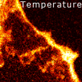
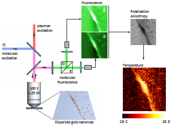 We report on a thermal imaging technique based on fluorescence polarization anisotropy measurements, which enables mapping the local temperature near nanometer-sized heat sources with 300 nm spatial resolution and a typical accuracy of 0.1°C. The principle is demonstrated by mapping the temperature landscape around plasmonic nano-structures heated by near-infrared light. By assessing directly the molecules' Brownian dynamics, it is shown that fluorescence polarization anisotropy is a robust and reliable method which overcomes the limitations of previous thermal imaging techniques. It opens new perspectives in medicine, nanoelectronics and nanofluidics where a control of temperature of a few degrees at the nanoscale is required.
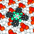
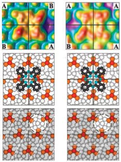 We report a description of the SiC(0001)3x3 silicon carbide reconstruction based on single molecule scanning tunneling microscopy (STM) observations and density functional theory calculations. We show that the SiC(0001)3x3 reconstruction can be described as contiguous domains of right and left chirality distributed at the nanoscale, which breaks the to date supposed translational invariance of the surface. While this surface heterochirality remains invisible in STM topographies of clean surfaces, individual metal-free phthalocyanine molecules chemisorbed on the surface act as molecular lenses to reveal the surface chirality in the STM topographies. This original method exemplifies the ability of STM to probe
atomic-scale structures in detail and provides a more complete vision of a frequently studied SiC reconstruction.
We report a description of the SiC(0001)3x3 silicon carbide reconstruction based on single molecule scanning tunneling microscopy (STM) observations and density functional theory calculations. We show that the SiC(0001)3x3 reconstruction can be described as contiguous domains of right and left chirality distributed at the nanoscale, which breaks the to date supposed translational invariance of the surface. While this surface heterochirality remains invisible in STM topographies of clean surfaces, individual metal-free phthalocyanine molecules chemisorbed on the surface act as molecular lenses to reveal the surface chirality in the STM topographies. This original method exemplifies the ability of STM to probe
atomic-scale structures in detail and provides a more complete vision of a frequently studied SiC reconstruction.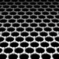
 Z-V scanning tunneling spectroscopy is used to probe the topology and electron scattering properties of the electronic interfaces of monolayer and bilayer graphenes, expitaxially grown on SiC(0001). The dZ/dV spectra validate existing calculations of the interface topology and provide evidence for new electron scattering properties due to changes in the electronic character of the bonding. Two sharp boundaries are observed: between the vacuum and the graphene pi state lying above the graphene atom plane and a subsurface barrier between the carbon-rich layer and the bulk SiC.
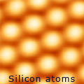
 We investigate charge transport through electronic surface states of the 6H-SiC(0001)-3×3 surface. Three intrinsic surface states are located within the wide bulk band gap, in which two (S1 and U1) arise from strongly correlated electronic states and the third (S2) has negligible electron correlation effects. Combined conductance and luminescence experiments with the scanning tunneling microscope show that the Mott-Hubbard surface states (S1 and U1) have a high resistance (1.0 GΩ), while the noncorrelated state (S2) has a negligible resistance. Consequently, current can be selectively transported through any of these three surface states.
 Molecular fluorescence decay is significantly modified when the emitting molecule is located near a plasmonic structure. When the lateral sizes of such structures are reduced to nanometer-scale cross sections, they can be used to accurately control and amplify the emission rate. In this Rapid Communication, we extend Green's dyadic method to quantitatively investigate both radiative and nonradiative decay channels experienced by a single fluorescent molecule confined in an adjustable dielectric-metal nanogap. The technique produces data in excellent agreement with current experimental work.
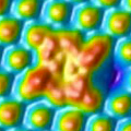
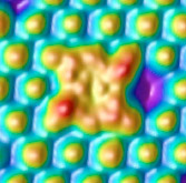 The adsorption of individual metal-free phthalocyanine molecules on the 6H-SiC(0001)3×3 surface was studied using the scanning tunneling microscope supported by density functional theory calculations. Phthalocyanine molecules were found to be chemisorbed through a reaction of two conjugated imide groups with two silicon adatoms. This type of anchoring opens numerous perspectives for the organic functionalization of a biocompatible wide band gap semiconductor.